Cervical disc herniation, lumbar disc herniation,
regenerative medicine disc herniation of spinal canal stenosis and cervical spondylotic myelopathy

  • HOME
  • Treatment
  • Regenerative medicine for cervical disc herniation, lumbar disc herniation, spinal canal stenosis, and cervical spondylotic myelopathy

The latest treatment technology called regenerative medicine (stem cell therapy)
Not conventional intravenous therapy
Direct intraspinal injection therapy

Our unique
treatment

Cervical disc herniation] Postoperative sequelae, numbness in limbs, and
difficulty in walking, treated with regenerative medicine, case report of a man in his 70s.

Lumbar disc herniation] Patient dramatically improved by stem cell therapy!
Before the treatment, I was scared and walked looking down...
But now I can walk looking forward! Here is a voice of joy!

What you can no longer do, you can do again.

Are you suffering from these symptoms
?

Latest treatment techniques to improve symptoms of cervical and lumbar hernias and spinal canal stenosis

If you are suffering from cervical disc herniation, lumbar disc herniation, spinal stenosis, or cervical spondylotic myelopathy, keep reading. The latest treatment technology called regenerative medicine (stem cell therapy) may be the answer to your problems.

We are able to offer the latest approach of regenerative medicine for the following conditions listed on the right.

We can provide regenerative medicine at our clinic
Hernia Symptoms
  • cervical disc herniation
  • lumbar disk herniation
  • spinal canal stenosis
  • cervical spondylotic myelopathy

First, let me explain each of these pathologies at
.

What is a herniated disc?

Theintervertebral disc consists of two layers of cartilage tissue called the nucleus pulposus (jelly-like nucleus pul posus that contains a lot of water) and the fibrous ring that surrounds the nucleus pulposus.

The spine moves flexibly and supports body weight by means of cushions called "intervertebral discs" between bones. However, due to aging or carrying heavy loads, the intervertebral discs can become deformed and pop out. This is a herniated disc.

Cervical disc herniation,
Lumbar disc herniation

Herniation in the cervical spine is called cervical disc herniation, and herniation in the lumbar spine is called lumbar disc herniation. This herniation causes numbness in the limbs, movement disorders, muscle weakness, and other symptoms.

A hernia is a protrusion of the internal medullary nucleus.

What is a herniated disc?

spinal canal stenosis

The spine has a tunnel called the spinal canal, through which the spinal nerves pass.

And on the back side of the spinal nerves is the yellow ligament. Spinal canal stenosis, which is common in middle-aged and older people, is a condition in which the tunnel is narrowed and the spinal canal is narrowed due to hernia protrusion, thickening of the yellow ligament, or deformation of the vertebral body itself.

This causes numbness in the limbs, movement disorders, and intermittent claudication (numbness in the buttocks and legs when walking, which becomes easier when resting).

Cervical disc herniation, lumbar disc herniation

cervical spondylotic myelopathy

Symptoms include numbness and pain in the hands, muscle weakness, and gait disturbance such as frequent stumbling while walking, due to compression of the spinal cord behind the cervical vertebrae caused by herniated disc protrusion, thickening of the yellow ligament, and deformation of the vertebra itself.

Dr. Sakamoto will explain! (Chairman of the Board)

Treatment with regenerative medicine
Who is suitable?

People like this one
are indicated for regenerative medicine.

Hernia, scoliosis, etc.

  • Numbness and
    pain in limbs but
    not indicated for surgery
    diagnosed
  • I have never had
    surgery yet
    I really want to
    avoid surgery
  • Unable to undergo surgery
  • I had surgery, but
    there are aftereffects.

I've had hernia and scoliosis surgeries.

  • Symptoms recurred.
  • I had surgery but my
    symptoms are still the same.
  • I had surgery.
    It got even worse.
  • Also in new locations
    Occurrence.

Surgery for cervical disc herniation, lumbar disc herniation, spinal canal stenosis, and cervical spondylotic myelopathy inevitably entails the risk of aftereffects due to the nature of the nerves involved. Even after surgery, numbness and pain may remain, and recovery of muscle strength may not be possible. On the contrary, symptoms that were not present in the first place may appear after surgery.

If you have a hernia

Have you heard of regenerative medicine?

Have you heard of regenerative medicine?

The clinic offers free consultations at

Free Consultation
  • Treatment methods and results
  • Unique stem cell therapy technology
  • Safety and risks
  • Treatment plans and pricing
Chairman Sakamoto
Call for a free consultation 0120-706-313 Toll free/ Office hours: 9:00-18:00

Do you have any
such
problems after surgery?

Painful aftereffects after surgery

After going through the trouble of surgery, symptoms such as numbness, pain, and muscle weakness have reappeared. Or, symptoms are worse than before the surgery, or symptoms that did not exist before the surgery have appeared after the surgery.

I had surgery but bladder rectal disorders such as frequent urination and constipation have not gone away. Or, symptoms of bladder rectal disorders that did not exist before the surgery have appeared.

Painful aftereffects after surgery Painful aftereffects after surgery
  • Numbness, pain, muscle weakness

  • Worsening of symptoms, appearance of symptoms that were not present before surgery

  • Frequent urination and constipation not cured

  • Appearance of cysto-rectal disturbances that were not present preoperatively

Why? Worse after surgery.

Although nerve compression is firmly removed by surgery, a certain percentage of patients experience symptoms that are not relieved as expected or, conversely, become more severe, or symptoms that were not present before surgery appear.

This is due to the nature of nerves, which, if compressed for a period of time, may not recover from the nerve damage or may gradually worsen thereafter, even after the compression that caused it is removed.

The longer the pressure is applied, the stronger the signs tend to be.

Why? Worse after surgery.

After surgery, too bad pronouncement

'The surgery was successful, there is no better way to treat it.'
'It's nerve surgery, so there will be some residual effects.'

When we talk to patients who come to our clinic, they are often told by their primary surgeon that "the surgery was successful and there is no further treatment" or "it is inevitable that there will be aftereffects because it is nerve surgery" and that they have to endure numbness and pain every day without other treatment options or that muscle weakness is interfering with their daily life. Most of them have to endure numbness and pain day after day without any other treatment.

Although various numbing and painkiller medications have been developed recently, it is often the case that symptoms are still not relieved at all.

I was told I had no choice.
Regenerative Medicine (Stem Cell Therapy) to improve the aftereffects

Our clinic is working on regenerative medicine to improve the aftereffects of surgery that was said to be unavoidable! Postoperative aftereffects are the limit of insurance treatment, and we can offer you a new option when you are told that there is nothing more you can do.

Regenerative medicine can improve the aftereffects
So there are new possibilities!

  • Conventional insurance treatment has its limitations.

    Conventional insurance treatment has its limitations.

    I can't help it.
    I can't help it.

  • New Options for Pushing the Limits

    New Options for Pushing the Limits

    Focus on the possibilities that regenerative medicine and stem cell therapy offer

Sadanori Sakamoto, Regenerative Medicine Specialist

Our regenerative medicine is a method rarely used in Japan called "direct intraspinal injection therapy," in which stem cells are administered directly into the spinal cord cavity for patients whose symptoms have not improved after surgery or have worsened further. The stem cells used are cultured using our clinic’s original method without freezing and then administered for treatment.

If you have a hernia

Have you heard of regenerative medicine?

Have you heard of regenerative medicine?

The clinic offers free consultations at

Free Consultation
  • Treatment methods and results
  • Unique stem cell therapy technology
  • Safety and risks
  • Treatment plans and pricing
Chairman Sakamoto
Call for a free consultation 0120-706-313 Toll free/ Office hours: 9:00-18:00

Our Strengths in Regenerative Medicine - Direct Intracavitary Spinal Cavity Injection Therapy -.

The key is direct into the spinal cord cavity!
Stem cells are more effective at regenerating and repairing nerves when they're administered directly into the spinal cord cavity.

Direct administration of stem cells to the damaged area Our unique intraspinal direct injection therapy!

With an IV, the numbers are down by the time
reaches the wrong part of the spinal cord.

The most common method of regenerative medicine for spinal cord injury is to administer stem cells intravenously and allow them to reach the spinal cord through the blood circulation. However, this method tends to limit the quantity of stem cells that can reach the injured spinal cord because stem cells injected into blood vessels are distributed throughout the body.

IV infusion reduces the number of stem cells reaching the trunk. IV infusion reduces the number of stem cells reaching the trunk.
Dr. Sakamoto, regenerative medicine specialist, our treatment is effective because it delivers stem cells directly to the affected area.

We offer a new treatment that has rarely been reported in Japan: the direct administration of stem cells to the damaged nerve site of the spinal cord. This treatment method directly administers stem cells to the damaged area of the spinal cord and is expected to repair and regenerate nerves near the spinal cord better than intravenous administration, which is effective throughout the body.

Intraspinal Direct Injection Therapy

Stem cell therapy administered directly into the spinal cord
STEP 1
It is administered by injection with a fine needle into the subarachnoid space (spinal cavity).
STEP2
After administration, stem cells flow into the spinal fluid.
STEP 3
Spinal fluid flows through the spinal cord and can deliver stem cells to the site of spinal cord injury.

The treatment is a simple injection that takes only a few minutes and is not very painful. Further effects can be expected when combined with stem cell infusion.

Because it's injected directly into the damaged area of the spinal cord,
it's more effective at regenerating and repairing the spinal nerves
!

Highly effective
direct intraspinal injection therapy
regenerative medicine

Generally, the only method of regenerative medicine for spinal nerves has been intravenous (intravenous) administration.

This conventional method cannot deliver a large number of stem cells into the spinal cord. In "Direct Intra-Spinal Cavity Injection Therapy," a large number of stem cells can directly regenerate the damaged nerves.

As a result, the effect is much higher than that of conventional infusion.

Highly effective intraspinal direct injection therapy regenerative medicine

Of course, intravenous stem cell therapy also has its advantages. It is possible to repair nerves that cannot be reached by "direct intraspinal injection therapy.

However, the significance of administering stem cells directly into the spinal cord cavity is very significant because most of the nerve damage in hernias and scoliosis is within the spinal cord.

Our clinic has submitted a notification to the Ministry of Health, Labour and Welfare for the treatment of "administration of stem cells into the spinal cord cavity," which is rarely performed in Japan, and it was accepted. 

Our clinic has received approval from the Ministry of Health, Labour and Welfare for the treatment of "administration of stem cells into the spinal cord," which is rarely done in Japan.

Let's take a look at an actual case at
.

case report

Dr. Sakamoto is also on YouTube! Check out his regenerative medicine channel. Dr. Sakamoto is also on YouTube! Check out his regenerative medicine channel.

[hernias, scoliosis, and myelopathy of the neck and lower back] A physician explains stem cell therapy for diseases that have damaged nerves in the spinal cord

Sadanori Sakamoto, Regenerative Medicine Specialist

In this way, regenerative medicine has been shown to improve symptoms that could not be helped by conventional treatments. Of course, there are individual differences, but the results are often astonishing, and I myself am often surprised by the results of the treatment. I hope to continue to provide cutting-edge regenerative medicine as a bridge to a ray of hope, so that people can do what they had given up on.

If you have a hernia

Have you heard of regenerative medicine?

Have you heard of regenerative medicine?

The clinic offers free consultations at

Free Consultation
  • Treatment methods and results
  • Unique stem cell therapy technology
  • Safety and risks
  • Treatment plans and pricing
Chairman Sakamoto
Call for a free consultation 0120-706-313 Toll free/ Office hours: 9:00-18:00

Our Stem Cell Strengths

Characteristics of our hospital (1)

A New Stage in Stem Cell Therapy
Regenerative Medicine Moves into the Era of Differentiation Induction|Joint Therapy

Regenerative medicine using differentiation induction enables more extensive repair of articular cartilage, meniscus, and osteonecrotic areas.

Delivering what you need where you need it (differentiation induction)
"The Next Generation of Regenerative Medicine"

A New Stage of Stem Cell Therapy
The Next Generation of "Regenerative Medicine " for Joints by Differentiation Induction

In our body, there are "stem cells," that can change into various forms like a fertilized egg.

For example, it can transform itself into nerves, blood vessels, bones, blood, muscles, etc.

This ability to transform is called differentiation..

I see that stem cells can differentiate into various tissues such as nerves, blood vessels, bone, blood, muscle, etc.

Stem cell differentiation

The process of guiding stem cells that change into various forms to differentiate into specific tissues such as bones and nerves is called "induction of differentiation.

By inducing differentiation, the administered stem cells can be fully utilized, thereby increasing the regenerative capacity of the targeted tissue.

By inducing stem cells to differentiate into
specific tissues, such as nerve or bone, you can increase the regenerative capacity of the
tissue you want to repair!

Induction of stem cell differentiation

Safety and Security

In this differentiation induction process, we do not use any chemical substances or manipulate genes. We use the power of our own cells, so you can receive our treatment with peace of mind.


regenerative medicine for joints by induction of differentiation

The cartilage of the joints has a foundation, which is called the subchondral bone (nankotsukakotsu).

By differentiating and inducing stem cells in this subchondral bone, it is now possible to prepare the foundation and generate a lot of cartilage on top of it, and also to maximize the effect on pain.

In order to regenerate cartilage in the joints, it is important to prepare the "subchondral bone,"
which is the foundation of the cartilage?

Regenerative medicine of joints by induction of differentiation

Characteristics of our hospital (2)

The quality of
stem cells is high because they are cultured without freezing.

Since the stem cells are cultured each time they are administered without freezing, they boast a high survival rate.

Our original culture, "as-is" culture

It's actually time-consuming to culture them "each time" as they are administered. But we use this method because it increases the survival rate at the time of administration!

Freshly cultured cells are fresh and lively! It is unique to our hospital that cells with high survival and activity rates can be administered every time.

Our original culture, "as-is" culture

As the cryopreservation period increases, the cells gradually die, and the surviving cells also lose quality.

Foods also lose their taste and quality when frozen!
It is best to use "freshly prepared" food made "at the time of use" for both cooking and cellular products!

Exactly! I would say that the quality of our unique "on-the-spot" culture is completely different from the general "make-ready" culture.

In Typical Cell Processing Facilities...
Cells are often frozen during storage and transport.

In addition, cryopreserved cells that are made and stored in cell processing rooms are generally transported frozen to other medical facilities.

These cryopreserved cells are transported to medical facilities while frozen, then thawed before use.However, many cells die or weaken during the thawing process, reducing their effectiveness.

General cell processing room General cell processing room

Features of our hospital (iii)

Using the patient's own blood
Safe culture method without blood substitutes

We do not use blood substitutes in cell culture.

I don't use it.
  • Blood substitutes
    (bovine serum, artificial serum)
We do not use blood substitutes in cell culture.

We insist on using only your own cells and
blood!

High safety and survival rates

At our clinic, we culture with your own blood, which is free of impurities and chemicals, ensuring high safety and survival rates. Some facilities use bovine blood or artificial blood called serum-free medium for culture.

High safety and survival rates

Generally, bovine blood and serum-free culture media are used for research purposes, but there are still concerns about allergies and mad cow disease in the clinical setting when actually administered to the human body.

In addition, although culturing stem cells from one's own blood requires advanced culturing techniques, stem cells cultured from one's own blood have strong vitality and can firmly repair damaged areas, enabling treatment with high regenerative potential.

High safety because cells are cultured using the patient's own blood

It is very difficult to culture them using their own blood, but they grow more vibrant cells that way.

I guess using your own cells and blood is more
compatible.

Features of our hospital (4)

Advanced technology for stem cell extraction
Proprietary separation sheets without chemicals or additives

We do not use chemicals or additives in cell culture

I don't use it.
  • Lipolytic enzymes
    (Chemicals)
  •  additive
We do not use any chemicals or additives during cell culture.

No chemicals or additives are used!

High safety and security /
Original culture technology

  • Using our own separation sheet, it is possible to culture stem cells with strong viability.
  • In culture methods using common degradative enzymes (chemicals), cells are weak and survival rates are low. Allergy is also a concern.
High safety and security / Original culture technology

Characteristics of our hospital (5)

High survival and activity rates

Stem cell viability and activity

Understanding stem cell viability and activity rates is very important in understanding regenerative medicine. Even when we talk about stem cell regenerative medicine, there is no uniformity in the way cells are cultured at different medical institutions. This means that the degree to which stem cells are viable and effective also varies from institution to institution.

In other words, the survival and activity rates of stem cells vary from institution to institution, and if the survival and activity rates are low, the desired effect cannot be expected.

Now we will discuss survival and activity rates.

What is Survival Rate?

Viability is the percentage of cultured stem cells that are alive.

Our Stem Cell Culture

Our stem cell culture, high survival rate

General culture

General culture, low survival rate

What is activity rate?

The activity rate is the percentage of how healthy the living stem cells are.

Our Stem Cell Culture

Our stem cell culture, high activity rate Our stem cell culture, high activity rate

General culture

General culture, low activity rate General culture, low activity rate

Even at the same survival rate, if the living cells are
weaker, the fixation rate will also be lower.

No matter how high the survival rate is, if the living cells are inactive and weak, the stem cells will not be fully effective. In other words, even if the survival rate is high, if there are many weak cells with a low activity rate, the stem cell therapy will be less effective, the stem cell fixation rate will be low, and the therapeutic outcome will be poor.

Features of our hospital (6)

The amount of fat to be extracted is small (about 2-3 grains of rice)
and therefore less stressful to the body

A 1 cm incision is made around the lower abdomen and fat (about 2-3 grains of rice) is harvested. Stem cells are extracted and cultured from the harvested fat cells, and the number of stem cells is increased to 10 to 100 million before they are administered into the body. (The number of cells administered varies depending on whether it is intravenous infusion, intra-articular injection, or intraspinal injection.)

Because stem cells are grown in culture, fewer cells are needed to be harvested, the wound is smaller, and there is almost no pain, so the burden on the body is minimal.

Less burden on the body due to the small amount of cells collected (2-3 grains of rice)

Characteristics of our hospital (7)

Because more than 100 million cells can be administered,
treatment results are good

Since the stem cells are cultured each time they are administered without freezing, they boast a high survival rate.

Comparison of treatment outcomes
(for cartilage regeneration)

General Treatment

  • BEFORE (before stem cell administration) Sakamoto: "The light brown area is bare bone." BEFORE (before stem cell administration) Sakamoto: "The light brown area is bare bone."
  • Stem cell administration: 10 million typical doses Stem cell administration: 10 million typical doses
  • AFTER (6th month of stem cell administration) Cell-kun: "The white area is newly regenerated cartilage!" AFTER (6th month of stem cell administration) Cell-kun: "The white area is newly regenerated cartilage!"

Our Treatment

  • BEFORE (before stem cell administration) Sakamoto: "The light brown area is bare bone." BEFORE (before stem cell administration) Sakamoto: "The light brown area is bare bone."
  • Stem cell administration: 100 million typical doses Stem cell administration: 100 million typical doses
  • AFTER (6th month of stem cell administration) Cell-kun: "The white area is newly regenerated cartilage!" AFTER (6th month of stem cell administration) Cell-kun: "The white area is newly regenerated cartilage!"

Detailed treatment information

Treatment details
Cell therapy using autologous adipose-derived mesenchymal stem cells (intra-articular knee injection).
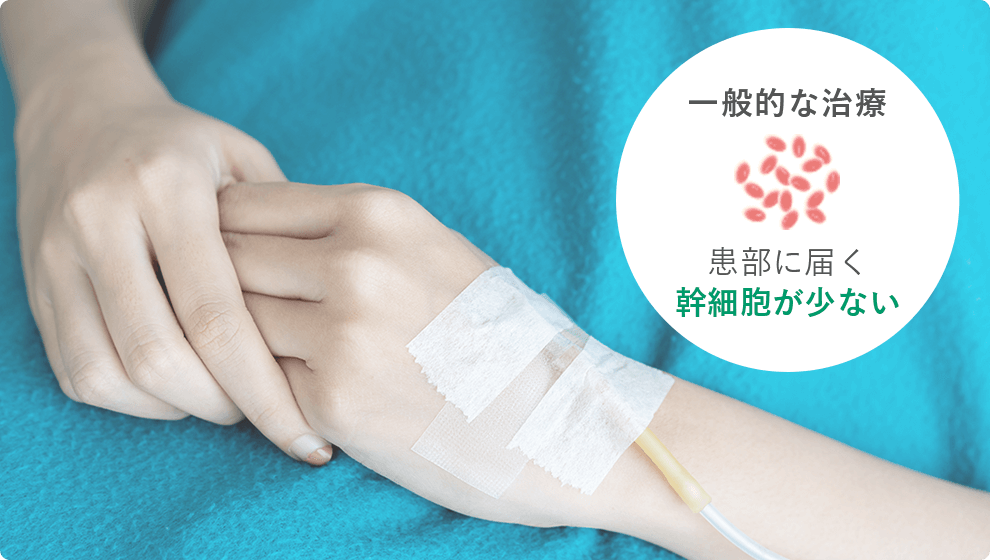
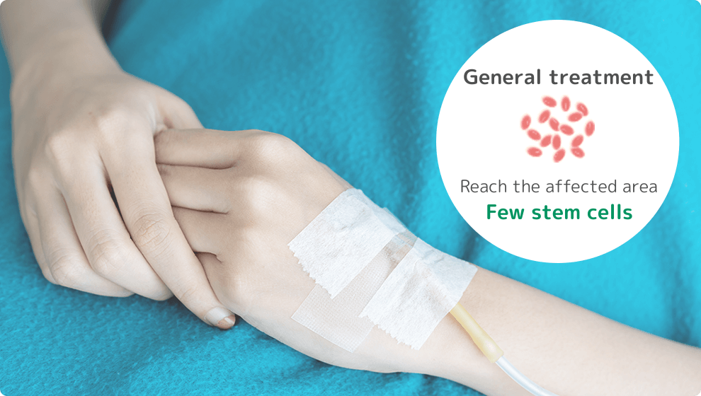
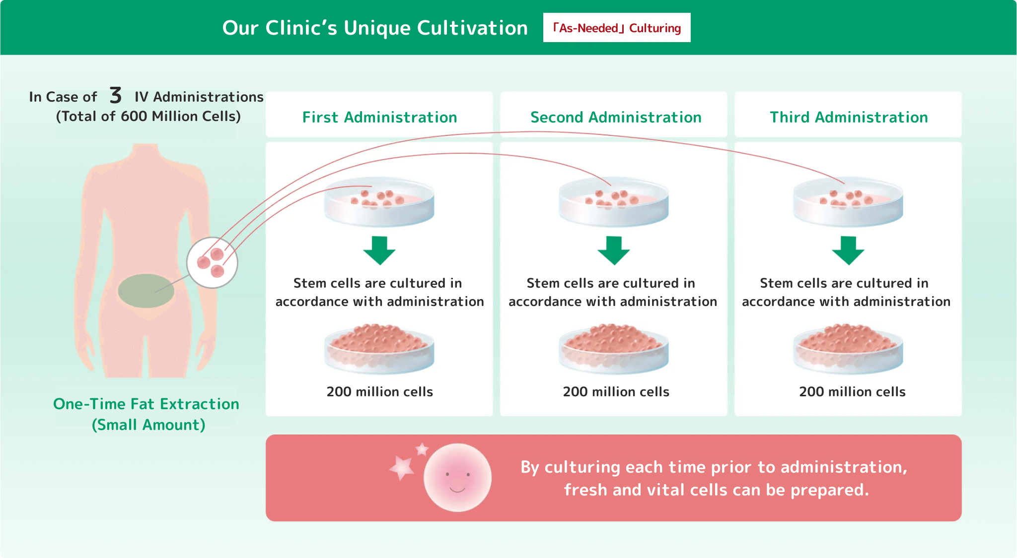
Since the cells are cultured each time they are administered without freezing, the stem cells have a high survival rate.

Duration and number of treatments

Studies have shown that effects begin to appear 3-6 months after administration.

cost

25 million pieces: 1,320,000 yen (including tax)
50 million pieces: 1,540,000 yen (tax included)
100 million pieces: 1,980,000 yen (tax included)
*This is an unreserved treatment.
A counseling fee of 3,300 yen (including tax) and a blood test fee of 11,000 yen (including tax) are required.
*The price is for a single dose. Discounts will be applied as the number of doses increases.

Major Risks and Side Effects

Pain, swelling, and internal bleeding at the treatment site (generally resolves in a few days)
Infection (extremely rare, but infection may occur during cell culture or administration)
Allergic reactions (extremely rare due to the autologous nature of the cells)
Individual differences in efficacy (efficacy may vary depending on symptoms and age)

The above image was taken from the article " Intra-articular injection of mesenchymal stem cells for the treatment of osteoarthritis of the knee: a proof-of-concept clinical trial. The image above is from the article "Intra-articular injection of mesenchymal stem cells for the treatment of osteoarthritis of the knee: a proof-of-concept clinical trial.
In this study, the best results were obtained in the high-dose (100 million stem cells) group. Effectiveness may vary from person to person. Please contact our clinic for more information.
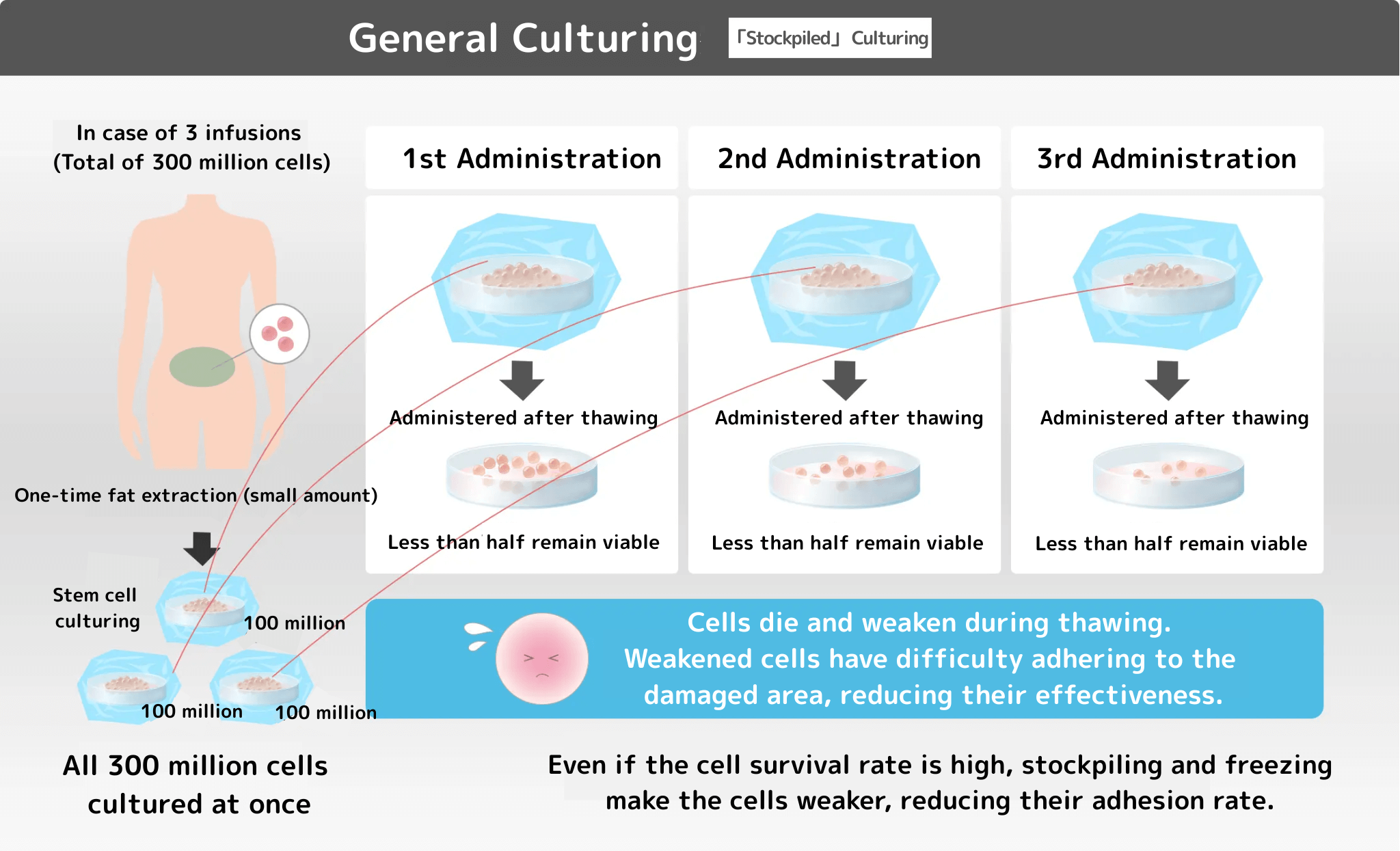
Compared to 10 million,
100 million stem cells administered is more
cartilage!

Conclusion. Conclusion. The more stem cells administered, the more
Good treatment results

In the picture, you can clearly see that the more stem cells administered, the more cartilage
is regenerated!

It is common practice to administer about 10 million stem cells in a joint. However, at our clinic, we can administer more than 100 million fresh stem cells that have not been cryopreserved, depending on the patient's symptoms.

It has been proven in clinical data from overseas that the greater the number of stem cells administered, the better the therapeutic outcome.

In addition..!

The quality assurance by a third-party organization makes
safe and secure.

All stem cells confirmed to meet quality standards

All stem cells confirmed to meet quality standards

Stem cells cultured under strict control are
evaluated for quality by a third party
to see how they really are!

It's like a certificate of authenticity that proves that the diamond ring is real and of what quality.

Stem Cell Therapy Procedure

At our clinic, stem cell therapy is performed according to the following procedure.

  • Initial consultation and interview (approx. 2 hours)

    01

    Initial consultation and interview (approx. 2 hours)

    The physician will conduct a detailed investigation of the patient's condition and confirm whether stem cell therapy is suitable. After confirmation, the specific treatment process and details are explained, and if consent is obtained, a blood sample is drawn to test for infections and other diseases. After that, we will ask the patient to submit a consent form and start the treatment.

  • Fat collection (about 30 minutes)

    02

    Fat collection (about 30 minutes)

    Local anesthesia is administered to the patient's lower abdomen, and about three grains of fatty tissue are extracted.

  • Cell Culture Process

    03

    Cell Culture Process

    We have submitted a notification to the Ministry of Health, Labor and Welfare and professionally process adipose tissue collected at an accepted cell culture center into the specific cellular products required for treatment.

  • Stem cells are administered directly into the spinal cord cavity

    04

    Stem cells directly into the
    spinal cord cavity

    -I.V. administration (about 50 minutes)
    -Local administration (from about 5 minutes)

If you have a hernia

Have you heard of regenerative medicine?

Have you heard of regenerative medicine?

The clinic offers free consultations at

Free Consultation
  • Treatment methods and results
  • Unique stem cell therapy technology
  • Safety and risks
  • Treatment plans and pricing
Chairman Sakamoto
Call for a free consultation 0120-706-313 Toll free/ Office hours: 9:00-18:00

summary

The cervical spine is composed of seven bones, with the first (annulus), second (axial), and seventh (longus) cervical vertebrae having a special shape. The third through sixth cervical vertebrae have a similar shape.

The annular and axial vertebrae are structurally very different from the other cervical vertebrae. The annular-axial vertebrae have a complex joint structure and play an important role in the flexion and rotation motion of the cervical spine. It has a range of motion of 25 degrees of flexion and extension at the annulus occipitalis joint and 80 degrees of rotation on both sides at the annular-axial joint. Between each vertebra is a shock-relieving cushion called the intervertebral disc, which connects the vertebrae to each other.

The spinal canal in the spine contains motor nerves that carry commands from the brain (central nervous system) to the extremities, and sensory nerves that carry sensory information (hot, painful, etc.) from various parts of the body to the brain, collectively called spinal nerves. The root of the nerves that branch off from the spinal nerves is called the nerve root.

Traditionally, the intervertebral disc retains elasticity, but when the tissue fails, the nucleus pulposus, which is housed in the disc, may push out and compress the spinal nerve or nerve root, and this condition is called a "herniation. When this herniation occurs in the cervical spine (cervical spinal nerve), it is called a cervical disc herniation.

symptoms

There are two main types of hernias, depending on the direction of protrusion. Since each type has different symptoms, diagnosis is important to establish a treatment plan.

When the spinal cord is compressed (myelopathy), the initial symptoms are numbness in the fingers. This is followed by the onset of fine motor deficits that make it difficult to use chopsticks, write, and perform other activities. Symptoms initially appear on one side of the body, but many cases eventually become bilateral. Eventually, a limp-like gait (spastic gait) develops, and symptoms of numbness extending to the toes and lower limbs, as well as urinary and defecation disorders, also appear.

When nerve roots are compressed (radiculopathy) many develop from neck pain (posterior neck, upper scapula, interscapular, and shoulder blade area). Nerve root disease due to herniation is characterized by a relatively acute onset and intense pain. It is generally accompanied by a delayed onset of upper extremity pain and numbness in the fingers. This numbness is often unilateral and rarely bilateral.

Pain and numbness may persist for several weeks, then may abate gradually, but dull pain and numbness often remain, with mild relief in a few weeks to a few months.

  • normal

    normal
    normal
  • cervical herniation

    cervical herniation
    cervical herniation

treatment

When symptoms of neuropraxis (neck pain and unilateral upper extremity pain) are the primary concern, conservative therapy is the first priority. Conservative therapy involves immobilization with a cervical collar, depending on the symptoms. Physical therapy, such as massage, stretching, and physical therapy to the neck and shoulder girdle area, may also be combined with oral medications (muscle relaxants, anti-inflammatory analgesics, etc.).

When symptoms of myelopathy (e.g., upper extremity pain and difficulty with dexterity) are the predominant symptoms, B vitamins are effective in nourishing the nerves. If severe pain persists, block injections (stellate ganglion block, brachial plexus block, nerve root block, etc.) may also be used to alleviate pain.

If symptoms do not improve with continued conservative therapy or if muscle weakness in the upper extremities does not improve, surgical therapy is indicated. If numbness and paralysis of the limbs on both sides of the body, muscle atrophy, difficulty with fine motor skills, and gait disturbance are observed, conservative therapy is often ineffective and an early decision must be made.

Conservative Therapy Rehabilitation

manual therapy

Massage, stretching, and joint mobilization are performed. In addition, muscle relaxation and limited range of motion in the surrounding area should be improved to the extent possible.

therapeutic exercise

Exercise therapy focuses on isometric ⦅ muscle strengthening⦆ exercises without moving the joint. Movement is performed with caution, as strong movement may cause neurologic symptoms.

Correction of posture and movement

Improvement of anterior cervical posture⦅If you have a ⦆cat posture⦅ with the head forward of the shoulder joint position, we will correct your posture by keeping the shoulder blades in the correct position.

Points to keep in mind in daily life

  • Avoid working in the same position for long periods of time (computer work, games, reading, etc.)
  • Avoid carrying heavy objects
  • Avoid vehicles that are bumpy (e.g., long bus rides)
  • Use a pillow that fits your body (height, softness, material, etc.)
  • Avoid strenuous sports (even non-intense exercise requires caution)
  • Maintain correct posture (avoid hunching and slouching)
  • Beware of falls on bicycles and motorcycles (falls can cause spinal cord injuries)

procedure

Although surgery is an option for both nerve root disease and myelopathy, there are different criteria for symptoms and duration for which surgery is indicated.

Neuropathy

Basically, conservative treatment is often chosen, but surgery is considered if there is no change in symptoms after 4-5 months of treatment.

Myelopathy

Because conservative treatment is often ineffective and can worsen symptoms, surgery may be chosen before paralysis and motor or sensory impairment progresses too rapidly.

Typical Surgery

There are two types of techniques: one performed under direct vision and the other using an endoscope. Anterior decompression and immobilization: A 5 cm skin incision is made on the anterolateral side of the neck, and the disc is removed to treat the area compressing the spinal cord and nerve roots while avoiding the trachea and esophagus.

The tissue compressing the nerve is removed from this space. This procedure removes the cause of the numbness and pain symptoms, but it can also cause instability, so artificial or autogenous bone grafts are used.

Posterior intervertebral foramen enlargement (vertebroplasty)

A skin incision of about 5 cm is made from the posterior neck and the cervical vertebral arch is removed (open window) to widen the nerve pathway and remove the hernia that is compressing the nerve root and spinal cord. This procedure can remove the cause of neurological symptoms such as pain and numbness.

Surgery using an endoscope

The following section also introduces the aforementioned discectomy and intervertebral foramen magnification procedures when performed endoscopically.

- Endoscopic cervical discectomy (MECD)

The skin incision is about 2 cm and the procedure is performed using an endoscope, allowing for a one-week hospital stay. Because the procedure is performed under an endoscope, there is little possibility of damage to adjacent vertebrae, which can occur in conventional anterior fusion surgery. Because the procedure is performed without detaching the muscles attached to the spine, spinal stability can be maintained.

- Endoscopic cervical foramen magnification (FECF)

Indications in cervical hernias are limited. (It is indicated for nerve root symptoms in the upper body.) Surgery is performed in the supine (back) or lateral (side) position. A skin incision of about 1 cm is made, and an endoscope and surgical equipment are inserted through the incision. As with MECD, there is little muscle detachment, so spinal stability can be maintained.

Postoperative

After surgery under direct vision, the patient may wear a cervical collar, but will be released from bed the next day and begin gait training with a walker. Normally, patients are discharged from the hospital about two weeks after surgery. However, if there are signs of gait disturbance before surgery, postoperative rehabilitation may require a longer period of time.

With MECD and FECF, there is no need to wear a cervical collar, the transition to rehabilitation is smooth, and the time to discharge is very short.

Complications

Surgery is performed to relieve pressure on the spinal cord and nerve roots, and to ensure stability and fixation of the cervical spine. In recent years, endoscopic rather than direct visualization has been increasingly used. Although the procedure is performed with great care and magnification of the nerves and blood vessels, there is a risk of complications as described below.

  • Injury to esophagus and carotid artery
  • Injury to the dura mater (tissue surrounding the spinal cord)
  • Leakage of cerebrospinal fluid contained in the dura mater from the body associated with injury to the dura mater, and meningitis secondary to such leakage
  • Spinal cord or nerve damage (nerve damage caused when shaving the cervical spine)
  • Spinal cord compression due to postoperative hematoma formation (risk of quadriplegia)
  • Damage to grafted bone
  • wound infection
  • Pain and numbness in the extracted bone area
  • Disability to raise the upper limb
  • DVT

summary

The lumbar spine is composed of five vertebrae and has the largest vertebral body among vertebrae, playing an important role as a support mechanism for the trunk. Each vertebra is connected by intervertebral discs, intervertebral joints, and various ligaments to form the spinal canal, within which the spinal cord, a bundle of nerves, is housed. In the second lumbar vertebra and below, the nerve is called the spinal cauda equina nerve because it is shaped like a horse's tail, and it branches to the left and right and is distributed around the lower limbs (legs) and pelvis. These nerve branches are called nerve roots, and each nerve root passes through a gap in the bone called the intervertebral foramen to the lower limb.

The structure of the intervertebral disc consists of a layered structure of fibrocartilage called the annulus fibrosus on the outside and a nucleus pulposus containing proteoglycans in the center. The nucleus pulposus is a gelatinous tissue that contains water and has elasticity, and it softens the impact on the vertebrae. The nucleus pulposus is a gelatinous tissue that contains water and elasticity, and it dampens the impact on the spine. However, when the fibrous rings become brittle, a tear occurs and the nucleus pulposus protrudes, compressing the nerves. This condition is called lumbar disc herniation.

Because intervertebral discs are prone to degenerative changes due to poor blood flow, a decrease in the amount of proteoglycans in the nucleus pulposus can result in a lumbar disc herniation. Bipedal walking is also thought to be a major cause of lumbar disc herniation, as it places a heavy burden on the lumbar spine.

Lumbar disc herniation is most common in people in their 50s, and is about twice as common in men as in women. It is also relatively rare in patients under 20 years of age and after 70 years of age.

symptoms

Persistent and recurrent acute low back pain (hiccups) in the lumbar to buttock region is noted, as well as pain and numbness in one lower extremity. These lower extremity symptoms depend on the direction of hernia protrusion. The cause of low back pain in herniation may be disc degeneration, intervertebral arthritis, nerve root irritation, or myofascial ligamentous

Symptoms of the lower extremities

Herniation in the upper lumbar spine often results in the appearance of neuralgia in the thigh, while herniation in the middle to lower lumbar spine often results in complaints of sciatica. These pains and numbness can be severe and may take several weeks to subside.

Characteristic neurological findings also appear depending on the nerve root that is compressed. For example, herniation between the third and fourth lumbar vertebrae (L3)/lumbar vertebrae (L4) results in loss of muscle strength to extend the knee joint due to L4 nerve root compression, while herniation between L4/5 results in loss of muscle strength to turn the toes up and the ankle joint up due to L5 nerve root compression. In herniation between the L5/1st sacral vertebra (S1), there is a loss of strength to bend the toes and lower the ankle joint.

Such neurological symptoms often occur in one lower extremity, but depending on the direction of the hernia protrusion, symptoms may appear in both lower extremities, or, although rare, urinary and defecation may be impaired, so follow-up is important. In addition, once nerve tissue is damaged, it is difficult to repair, and neurological symptoms often remain even after treatment.

Treatment Options

Conservative therapy is the treatment of choice for those with mild or early symptoms, and about 80% of patients experience relief. Conservative therapy includes wearing a corset to maintain rest and physical therapy such as massage and stretching.

If the pain is severe, epidural block injections in the sacral or lumbar region are also used for pain relief, and oral medications such as muscle relaxants, anti-inflammatory analgesics, and vitamin B drugs are used in conjunction with these treatments.

Surgical treatment is selected when conservative treatment is ineffective for several months, the pain is frequent or increasing, motor paralysis of the lower limbs is pronounced, and urinary and bowel movements are disturbed.

Rehabilitation in Conservative Therapy

Massage, stretching, and joint range-of-motion exercises are performed without strain, taking care not to cause pain or numbness. Exercise therapy such as abdominal breathing and pelvic exercises are also effective.

abdominal breathing

  • This is strength training that can be performed without exercising the joints.
  • For those with pain or numbness, start with low-intensity training, as strong strength training may worsen symptoms.
  • Abdominal breathing increases abdominal pressure to maintain and improve spinal stability.

pelvic exercises

  • Sitting on a soft cushion and moving the pelvis back and forth improves lumbar spine and pelvic movement.
  • For those with hernias, pain and numbness cause less movement of the pelvis and lumbar spine, so it is important to expand the movement of the spine and pelvis as well as the muscles. The above exercises are just examples, and rehabilitation should be tailored to each individual's symptoms.

Points to keep in mind in daily life

  • Working in the middle of the back
  • Bending the upper body to lift heavy objects (when holding a heavy object, bend the knees together to lift the load).
  • Same posture for long periods of time (even sitting posture can strain the lower back over a long period of time)
  • Sudden getting up motion

Type of Surgery

Current surgical treatment for lumbar disc herniation depends on the symptoms and other factors, and the surgical technique chosen. Here we would like to introduce some of the typical surgical methods.

discectomy

- Lumbar discectomy (LOVE method)

This is a long-established technique for lumbar disc herniation. A 5 to 10 cm skin incision is made from the back side, the muscle is detached, and the vertebral arch is opened (a portion of the spine is opened). The nucleus pulposus and ring of fibers that are pressing on the nerves are then removed from this open area.

Because the surgery is performed under direct vision, lesions are less likely to be missed. There is also another type of surgery in which a microscope or endoscope is used to perform the same procedure, and the choice of surgical technique depends on the case. The hospitalization period is about 2 to 3 weeks.

discectomy

The MD and MED methods are the same techniques as the LOVE method with smaller skin incisions.

- Microscopic discectomy (MD procedure)

The skin incision is narrower than that of the LOVE method, at about 3 cm, and less invasive to the body because there is less muscle dissection. It is indicated for patients with moderate to severe lumbar spinal canal stenosis or lumbar spondylolisthesis. Hospitalization takes one to two weeks.

- Endoscopic discectomy (MED method)

The skin incision is about 2 cm in size, and after the incision is made, an endoscope is inserted to remove the protruding hernia while viewing the screen on a monitor. Because the skin incision is small, there are no stitches to be removed after the surgery, and there is little risk of infection. This procedure is also indicated for patients with moderate to severe hernias. Patients are able to walk the day after surgery and are hospitalized for approximately one week.

- Percutaneous endoscopic lumbar discectomy (PELD)

The skin incision is even narrower than in the MED method, and the hernia is removed using a 6mm operating tube and an endoscope or 3mm instrument. This technique is less invasive to the body with little or no muscle or ligament cutting, and there is no need for suture removal. Patients are able to walk within a few hours after surgery and can leave the hospital the same day or the next day. This treatment is not covered by insurance, so all costs are to be borne by the patient.

Other treatments

- Intradiscal enzyme injection therapy (Hernicore injection)

This is a treatment method to reduce nerve compression by injecting an enzyme-containing drug into the disc where herniation has occurred. Because it is performed under local anesthesia, the treatment can be performed as a day trip or overnight stay in the hospital.

In recent years, advances in medical care have led to an increase in the number of hospitals that perform surgeries that are not covered by insurance (e.g., laser treatment), expanding the range of options available.

Postoperative Progress

The following is an overview of the postoperative course of a typical surgical procedure. The patient may be fitted with a lumbar corset, but is released from bed the day after surgery and begins gait training with a walker. Although lumbar pain may persist for a few days, the patient does not need to rest any longer than necessary and is usually discharged 7-14 days after the surgery. After discharge from the hospital, patients continue to be monitored for neurological findings and diagnostic imaging such as X-rays and MRIs.

Postoperative hospital visits are usually required for about one year. Return to work or school depends on preoperative symptoms, but usually takes 1-2 months after surgery.

Surgical Complications

The goal of surgery is to relieve pressure on nerve roots and the spinal cord. The majority of surgeries currently performed in clinical practice use endoscopes or microscopes, which provide a bright, wide surgical field and magnify nerves and blood vessels for careful surgical procedures. However, depending on local conditions and other factors, postoperative complications and sequelae may occur.

Possible complications and sequelae

  • Paralysis or loss of motor and sensory function in the lower extremities due to nerve damage
  • Vesico-rectal disorders (disorders related to urination and defecation) due to nerve damage
  • Discitis with wound infection and inflammation
  • Spinal fluid leakage and secondary meningitis due to damage to the dura mater (membrane surrounding the nerves)
  • Nerve palsy and leg pain due to wound hematoma formation
  • Bleeding due to vascular injury in the abdomen
  • DVT
  • Pneumonia and other infectious diseases

summary

Spinal canal stenosis is a condition in which the spinal canal is narrowed due to osteophyte formation at the posterior margin of the vertebral body, thickening of the yellow ligament, or degeneration of the spine. Spinal canal narrowing can also be caused by spondylolisthesis (misalignment of the upper and lower spine). The most common sites of spinal canal stenosis are the cervical and lumbar vertebrae.

symptoms

cervical spinal canal stenosis

Symptoms appear due to several compression factors on the spinal nerves (degeneration of the spine and intervertebral discs and thickening of ligaments) and circulatory disturbances (inadequate circulation of blood flow to the spinal cord). Symptoms also vary depending on where the stenosis is found, and the speed of progression varies from person to person. Depending on the location of the nerve stenosis, the disease is classified into spinal cord type and nerve root type.

- 1. spinal cord type

The spinal nerves are compressed, resulting in neck pain, pain and numbness in both upper extremities, difficulty with fine motor skills, bladder rectal problems (difficulty with urination and defecation), and gait disturbance.

- 2. nerve root type

Nerve roots are compressed, causing neck pain, back pain, unilateral upper extremity pain and numbness. Initial symptoms begin with pain and stiffness in the neck and shoulder blades, followed gradually by pain with movement. As the spinal canal narrowing progresses, nerve roots are affected and pain and numbness appear in the neck, shoulder back, and upper extremities. Further narrowing of the spinal cord results in bilateral upper and lower extremity pain and numbness, and impaired fine motor skills.

If symptoms progress quickly or are extensive, bladder rectal problems and gait disturbance may occur, which can have a significant impact on daily life. The location of the radiating pain to the upper extremities can also be used to infer the nerve root(s) involved.

- Disrupted nerve root and the site of pain onset

  • C2 nerve root: occipital and temporal
  • C3 nerve root: occiput, behind the eye, behind the ear
  • C4 nerve root: posterior neck and upper scapula
  • C5 nerve root: upper scapula and lateral humerus
  • C6 nerve root: upper scapula and lateral humerus
  • C7 nerve root: interscapular, scapular, posterior part of upper limb
  • C8 nerve root: interscapular, scapular, medial humerus

lumbar spinal canal stenosis

Depending on the site of the nerve constriction, it is classified as cauda equina, nerve root, or mixed type.

- 1. cauda equina type

The central part of the spinal canal (cauda equina nerve) is compressed, causing bilateral leg pain, numbness, and bladder rectal problems.

- 2. nerve root type

The nerve root at the end of the branch from the cauda equina nerve is compressed. Pain occurs from the buttock to the lower extremity on one side.

- 3. mixed type

It is accompanied by symptoms of both the cauda equina type and the nerve root type.

Common symptoms include pain, numbness, and heaviness from the back of the lumbar region and buttocks to the posterior lower legs, calves, and feet, and patients rarely complain of pain in the lower back, even when spinal canal stenosis occurs in the lumbar region.

The nerves and surrounding blood vessels are also compressed, resulting in poor blood circulation in the spinal canal and the characteristic symptom of "intermittent claudication," in which walking becomes difficult due to pain and numbness in the lower limbs after a short walk, but walking can be resumed after a break.

Pain and numbness in the lower extremities when walking are relieved by bending forward, which releases the strangulated condition of the blood vessels and nerves and lightens the symptoms. Therefore, many patients are able to walk easily when pushing a silver car or shopping cart, and many have no problem riding a bicycle.

The nerve root that is being affected can also be inferred by the location of the radiating pain to the lower extremity.

- Disrupted nerve root and the site of pain onset

  • L2 nerve root: proximal femur
  • L3 nerve root: distal femur to knee joint area
  • L4 nerve root: medial leg
  • L5 nerve root: Lateral lower leg to toe (metatarsal to 2 toes)
  • S1 nerve root: lateral foot to lateral toe (3 to 5 toes)

Lumbar spinal stenosis is often contrasted with lumbar disc herniation, which has the characteristic symptom of intermittent claudication. Another difference is that herniated discs often develop acutely, whereas spinal canal stenosis progresses over a longer period of time.

treatment

Depending on the intensity of symptoms, conservative therapy (physical therapy, medication, block injections) is often the first choice.

physical therapy

Massage, stretching, electrotherapy, treatment with other physical therapy equipment, etc.

medical treatment by internal medicine

Common painkillers such as Loxonin and Voltaren Lyrica, Cymbalta, and other nerve pain medications Opalmon and other medications that improve blood flow

injection therapy

The cervical spine is treated with stellate ganglion block, brachial plexus block, and nerve root block. The lumbar spine is treated with sacral epidural block, lumbar epidural block, etc. They are effective in reducing pain, but are not permanent.

surgical treatment

If conservative treatment is not effective, surgery is considered. The decision to perform surgery is considered when the pain is so severe that it significantly interferes with daily life or when the distance that can be walked is reduced.

Early consultation with a doctor is also indicated for surgery if there is a possibility of residual aftereffects if left untreated or if there is a "bladder rectal disorder," so please consult with a doctor as soon as possible. Although the decision for surgery is difficult to make, the longer the disease has been present, the less likely it is to improve, so care should be taken in monitoring the progress of the disease.

surgical treatment

Although the surgical procedures for the cervical and lumbar spine differ slightly, the purpose of both is the same. The two basic methods are to remove degenerated bone and thickened ligaments that cause narrowing of the spinal canal and to fix the spine that has become unstable due to significant deformity. The following is an introduction to each of these surgeries.

cervical spinal canal stenosis

For cervical spine surgery, either an anterior or posterior approach is used.

- 1. anterior decompression and fusion of the cervical spine

The patient is placed in the supine position (on his/her back) for the surgery to be performed from the anterior neck. A skin incision is made from the anterior cervical region, and a portion of the cervical vertebrae is removed while avoiding the trachea and esophagus, and then proceeding to the spinal cord. After decompression to relieve nerve compression, an artificial device is inserted. The surgery is performed under general anesthesia and takes about one hour per vertebra. The hospitalization period is about 2 weeks.

- Endoscopic cervical vertebroplasty

The surgery is performed under general anesthesia in the supine position. A skin incision of less than 20 mm is made from the back of the neck, and the surgery is performed using an endoscope. Bones and ligaments that are pressing on the nerves are removed. The scar is small and there is minimal damage to the surrounding muscles, making this a less invasive procedure. The surgery takes about one hour and requires one week of hospitalization.

- Cervical vertebroplasty

If the stenosis is extensive, an endoscope cannot be used, and a direct visualization procedure may be chosen. The patient is placed in a supine position under general anesthesia. A skin incision is made from the posterior neck, and the muscles attached to the posterior cervical vertebral arch are removed. The vertebral arch on one side is separated, and an incision is made in the other vertebral arch to open it. An artificial bone is inserted into the open gap to remove the nerve compression. The surgery takes about one hour and requires approximately 10 days of hospitalization.

- Postoperative Progress

A cervical collar is worn postoperatively, but walking is possible from the next day. Postoperative rehabilitation may be longer for those who had difficulty walking before the surgery. After discharge from the hospital, patients will need to be examined every two weeks for a follow-up of about three months.

lumbar spinal canal stenosis

In the past, surgeries were typically performed under direct vision with a large surgical field, but now endoscopic surgeries are becoming the mainstream, so we will also introduce surgeries using an endoscope.

- 1. spondylolistectomy

The procedure is performed under general anesthesia and direct vision from the back. The extent of the skin incision depends on the affected vertebrae. First, the supraspinous ligament is dissected from the spinous process of the lumbar spine, followed by dissection of the spinal muscles. The vertebral arch is removed to relieve pressure on the spinal cord and nerve roots in the spinal canal.

- 2. window opening

This procedure is performed under general anesthesia and under direct vision from the back. The extent of the skin incision varies depending on the affected vertebrae, but the supraspinous ligament is preserved, the bilateral lumbosacral fascia are incised, and the spinal muscles are dissected from the sides of the spinous processes. The posterior portion of the vertebral arch is opened to relieve pressure on the spinal cord and nerve roots.

- 3. posterolateral lumbar fusion (PLF)

The patient is placed in a prone position under general anesthesia. A median incision is made from the spinous process of one vertebra above the affected vertebra to the lower vertebra to be fixed, and the supraspinous and interspinous ligaments are spared and the spinal muscles are dissected. The intervertebral joints, transverse processes, and soft tissues exposed by the dissection are resected to relieve pressure. Fixation after resection is performed with either grafted bone or metal fixtures.

- 4. posterior lumbar interbody fusion (PLIF)

The patient is placed in a prone position under general anesthesia. A 10 cm median incision is made from the back around the affected vertebra. The supraspinous and interspinous ligaments are preserved, the right and left vertebral arches and intervertebral joints are exposed, and a laminectomy and opening of the intervertebral foramen are performed. After slow pressure is applied, intervertebral spacers are inserted and fixed with spinal fusion hardware.

- Endoscopic lumbar laminectomy (MEL)

The procedure is performed under general anesthesia using an endoscope. A skin incision of about 2 cm is made on the back and the procedure is performed. The vertebral arch is resected and the thickened yellow ligament is removed to relieve nerve compression.

- Postoperative Progress

After surgery, the patient may wear a lumbar corset, but will begin gait training the day after surgery. Depending on the surgical technique, patients are often discharged from the hospital 7 to 14 days after surgery. Postoperative follow-up will be done by 3-12 month follow-up visits. Patients who have undergone fusion surgery must avoid excessive back and forth bending or twisting of the hips. It takes about six months for the bones to completely fuse, so it is necessary to live carefully.

Complications in surgery

The goal of cervical spine surgery is to relieve pressure on the spinal cord and nerve roots, while the goal of lumbar spine surgery is to relieve pressure on the nerves by widening the space for the spinal cauda equina nerve. Although the majority of surgeries are performed carefully using endoscopes and microscopes, under a bright surgical field and with great magnification of nerves, blood vessels, and other tissues, there is a great risk of complications arising from intraoperative manipulations. There is also a risk of complications arising from non-surgical procedural issues. The risk of the following complications is present in both cervical and lumbar spine surgeries, and any uncertainties should be confirmed prior to surgery.

  • Paralysis of the lower limbs due to nerve damage, paresthesia of the lower limbs, urinary and defecation disorders
  • Postoperative discitis with wound infection or inflammation of the disc
  • Leakage of spinal fluid due to damage to the membrane surrounding the nerves (dura mater) and associated meningitis
  • Nerve paralysis due to wound hematoma formation
  • Intraoperative major bleeding due to injury of large blood vessels in the lower extremities and abdomen
  • Infectious diseases such as deep vein thrombosis and pneumonia
  • Nerve damage from high speed rotary drills used to remove cervical vertebrae
  • Spinal cord compression due to postoperative hematoma formation
  • Prolapse or fracture of grafted bone
  • Wound infection
  • Appearance of pain and numbness in the extracted bone area
  • Stubborn pain in the neck
  • Deterioration of cervical spine balance
  • Disability to raise the upper limb
  • Difficulty swallowing or speaking
  • airway obstruction

summary

The spine is composed of 7 cervical vertebrae, 12 thoracic vertebrae, 5 lumbar vertebrae, sacral vertebrae, and tailbone. Spondylolisthesis is a disease in which the vertebrae that make up the spinal column (spine) slide back and forth, causing pain and numbness in the lower back and lower extremities. Normally, the spine does not slip easily, but when the spine, intervertebral joints, or intervertebral discs become unstable due to degeneration or damage, spinal slippage occurs. This spondylolisthesis often occurs in the lumbar spine.

preferred site

Often seen at L3-4, L4-5, and L5-S1, with L5-S1 being the most common occurrence. *L=lumbar spine/S=sacral spine

cause

It is caused by the loss of spinal stability due to degeneration of the intervertebral discs, ligaments, and joints caused by the strain of sports activities in youth and long-standing lifestyle habits.

Classification.

Typically, there are two types of slip due to degeneration and separation. In addition, although most slip is basically anterior, posterior slippage may also be seen.

degenerative slipping disease

It is most commonly seen in the elderly. It occurs when the stability of the upper and lower vertebrae is disrupted due to degeneration of the intervertebral discs caused by aging, laxity of the ligaments around the intervertebral joints, muscle weakness, and other factors.

spondylolisthesis

It is more common in young people. Excessive repetitive strain on the spine caused by sports and other activities can lead to the separation of a part of the spine, and as the condition progresses, the condition can shift to spondylolisthesis.

Spondylolisthesis is staged according to the degree of deviation from the adjacent lower vertebra. However, the actual symptoms must be taken into consideration when formulating a treatment plan.

Meyerding Classification

  • Grade I: 0-25%.
  • Grade II: 25 to 50%.
  • Grade III: 50-75%.
  • Grade IV: 75-100%.

symptoms

  •  lumbago
  •  Lower extremity symptoms (numbness, pain, muscle weakness, dysesthesia)
  •  intermittent claudication
  •  cysto-rectal obstruction

Spondylolisthesis presents similar symptoms to scoliosis.

The most common movement that causes lower back pain is bending forward. As the degree of slippage increases, narrowing of the spinal canal can occur, causing compression of the nerves (cauda equina and nerve roots), which can lead to symptoms of intermittent claudication.

Intermittent claudication is characterized by pain in the lower extremities after walking for a while, making it difficult to continue walking. However, after a short rest, the patient is able to walk again. Walking may also become difficult due to muscle weakness or sensory abnormalities.

diagnosis

image diagnosis

To check for spinal slippage on X-ray (x-ray) examination, the patient is placed in a forward (bending forward) and backward (bending backward) position and photographed from the side.

MRI scan

An MRI scan is used to determine the degree of nerve compression. It is useful when spinal cord or nerve root abnormalities are suspected.

manual checking

SLR test = lower extremity extension elevation test
FNS test = femoral nerve stretch test
SLR and FNS tests confirm the presence of nerve root involvement.

medical treatment

conservative therapy

  • Resting the affected area (wearing a corset)
  • Internal analgesic
  • Injection to remove inflammation
  • Muscle strengthening (strengthening abdominal, back, and gluteal muscles, etc.)
  • Increased flexibility (stretching of gluteal muscles and lower extremities)

When exercising a person with a slip, it is advisable to avoid movements that strongly warp or twist the spine.

Surgery is indicated when conservative therapy fails to produce changes. Surgery should also be considered when symptoms (pain and numbness in the lower extremities, gait disturbance, intermittent claudication, cysto-rectal disturbances, etc.) are severe and significantly affect daily life.

surgical treatment

Fusion is often used as a typical surgical procedure. In fusion surgery, the slipped spine is shaved to widen the nerve pathways and fixed using artificial or autogenous bone or metal. The extent of the skin incision and the method of fixation differ depending on the surgical technique, and the procedure is selected according to the individual's condition. If the degree of slippage is not advanced, decompression surgery may be performed to relieve pressure on the nerve.

Surgical Procedure

stereotomy

- PLIF (posterior intervertebral fusion)

A skin incision is made in the midline of the lumbar back, the spinous process is removed, and even part of the vertebral arch is resected. The intervertebral disc is removed while protecting the spinal cord to avoid intraoperative nerve damage. Autologous or artificial bone is inserted at the resection site and fixed with metal.

- TLIF (intervertebral fusion with unilateral entry)

The intervertebral joints on either side are removed, and the disc and bone spurs of the vertebral body are removed. Then, an artificial material is inserted into the removed disc and the vertebral body is fixed. Since only one side of the disc is removed, the bone can be preserved. Another advantage is that there is relatively little bleeding.

decompression surgery

- discectomy

A surgical procedure in which part of the vertebral arch and the yellow ligament are removed to relieve pressure on the spinal cord and nerve roots.

Postoperative course

From the next day, the patient wears a corset and undergoes rehabilitation according to his/her physical condition and pain. The rehabilitation includes exercises for getting up and down, gait training, etc. The average length of hospital stay is about 3 to 4 weeks. After discharge from the hospital, patients will continue to recuperate for about 2 weeks and return to their daily lives about 1 to 2 months after the surgery.

Postoperative Complications

  • infectious disease
  • Meningitis due to dural injury
  • Thrombophlebitis of the lower extremities
  • pulmonary embolism
  • nerve damage
  • Fixture Issues
  • Prolapse or fracture of grafted bone
  • Spinal cord compression due to postoperative hematoma formation
  • Intraoperative vascular injury

Regenerative medicine holds new possibilities. Through stem cell therapy, we can achieve what has been considered difficult.

We sincerely hope that this treatment will become more widespread and offer new hope to many people.

Medical institutions notified by the Ministry of Health, Labor and Welfare

Type II and Type III regenerative medicine, etc.
Plan of provision Notified

Repair Cell Clinic has submitted a Type 2 and Type 3 Regenerative Medicine Provision Plan to
Ministry of Health, Labour and Welfare and it has been accepted.

  • Ministry of Health, Labour and Welfare, Ordinance for Enforcement of the Act on Safety Assurance of Regenerative Medicine, etc., Notification Document "Treatment of Spinal Cord Injury Using Autologous Fat-derived Stem Cells".
  • Ministry of Health, Labour and Welfare, Ordinance for Enforcement of the Act on Safety Assurance of Regenerative Medicine, etc., Notification Document "Treatment of Spinal Cord Injury Using Autologous Fat-derived Stem Cells".

Using autologous adipose-derived stem cells to treat
spinal cord injury

Our clinic provides free treatment under the Law for Ensuring the Safety of Regenerative Medicine for the Treatment of Spinal Cord Injury and other cutting-edge regenerative medicine using autologous stem cells, which are few in Japan, and intra-articular administration of PRP (platelet-rich plasma). The Ministry of Health, Labour and Welfare (MHLW) has approved regenerative medicine, which is subject to strict review by the Specified Authorized Committee for Regenerative Medicine, and only when the appropriateness, safety, and physician system of the treatment are recognized as appropriate can a treatment plan be submitted to the MHLW.

Repair Cell Clinic for Regenerative Medicine TOPtop